Bursitis Of The Foot Bursa Removal Complications
Overview
Achilles tendon bursitis occurs mainly in young women but can develop in men. Walking in a way that repeatedly presses the soft tissue behind the heel against the stiff back support of a shoe can cause or aggravate the bursitis. Shoes that taper sharply inward toward the posterior heel (such as high-heeled shoes) can cause irritating pressure that leads to the development of this bursitis. Normally, only one bursa is in the heel, between the Achilles tendon and the heel bone (calcaneus). This bursa may become inflamed, swollen, and painful, resulting in anterior Achilles tendon bursitis. Abnormal pressure and foot dysfunction can cause a protective bursa to form between the Achilles tendon and the skin. This bursa may also become inflamed, swollen, and painful, resulting in posterior Achilles tendon bursitis. Any condition that puts extra strain on the Achilles tendon can cause anterior Achilles tendon bursitis. Injuries to the heel and diseases such as rheumatoid arthritis can also cause it.
Causes
Age. Bursitis is more common during middle age due to repetitive activities that put wear and tear on the body over time. Certain activities or occupations. If your job or hobby involves repetitive motion or puts pressure on bursae, you have a higher likelihood of developing bursitis. Reaching overhead, leaning elbows on arm rests, crossing your legs, laying carpet, setting tile, gardening, biking, playing baseball and ice skating are some activities that, when repeated very often, can put you at increased risk of developing bursitis. Sports in which you may get hit in the knee or fall to the knees, such as football, can also increase the risk. Some medical or health conditions. Rheumatoid arthritis, osteoarthritis, gout, thyroid disease, diabetes, alcoholism and some immunosuppressive disorders can increase the risk of bursitis. The reasons can vary, from cartilage breakdown around joints (arthritis) to crystals in the bursa that cause inflammation (gout). Wearing high heels. Posterior Achilles tendon bursitis occurs when the bursa located between the skin and the Achilles tendon (the band of tissue that attaches the calf muscle to the heel bone) becomes inflamed. High heels are often to blame for this, the stiff heel can put direct pressure on the bursa between the skin and the Achilles tendon.
Symptoms
What are the symptoms of heel bursitis? pain, swelling, tenderness, redness, and/or warmth at either the bottom of the heel or top of the heel, depending on the degree of swelling, pain may be a dull ache or substantial enough to cause limping, running, jumping, and walking activities may exacerbate pain, wearing poorly fitting, tight, or high-heeled shoes may exacerbate pain.
Diagnosis
A good clinical practise includes evaluation of the tendon, bursa and calcaneum by, careful history, inspection of the region for bony prominence and local swelling as well as palpation of the area of maximal tenderness. Biomechanical abnormalities, joint stiffness and proximal soft tissue tightening can exacerbate an anatomical predisposition to retrocalcaneal bursitis, they warrant correction when present.
Non Surgical Treatment
Physiotherapy treatment is vital to hasten the healing process, ensure an optimal outcome and reduce the likelihood of injury recurrence in all patients with retrocalcaneal bursitis. Treatment may comprise soft tissue massage (particularly to the calf muscles), joint mobilization (of the ankle, subtalar joint and foot), dry needling, electrotherapy (e.g. ultrasound), stretches, the use of heel wedges, the use of crutches, ice or heat treatment, arch support taping, the use of a compression bandage, exercises to improve strength, flexibility, balance and core stability, education, anti-inflammatory advice, activity modification advice, biomechanical correction (e.g. the use of orthotics), footwear advice, a gradual return to activity program.
Surgical Treatment
Surgery is rarely need to treat most of these conditions. A patient with a soft tissue rheumatic syndrome may need surgery, however, if problems persist and other treatment methods do not help symptoms.
Hammer Toe Concerns
 Overview
A Hammer toe is a toe that is bent because of a weakened muscle. The weakened muscle makes the tendons (tissues that connect muscles to bone) shorter, causing the toes to curl under the feet. Hammertoes can run in families. They can also be caused by shoes that are too short. Hammertoes can cause problems with walking and can lead to other foot problems, such as blisters, calluses, and sores. Splinting and corrective footwear can help in treating hammertoes. In severe cases, surgery to straighten the toe may be necessary.
Causes
This condition is greatly influenced by the footwear we choose. Ladies who wear high heels are a perfect example. High heels force the toes to overlap and bend at the middle joint of the toe, resulting in hammertoe. But high heels are not the only culprits. Anyone who wears shoes that are too tight is increasing their risk of developing hammertoe. This progressive condition, which will only get better with treatment, can cause pain as the toes are forced to bend unnaturally.
 Symptoms
People with a hammer toe will often find that a corn or callus will develop on the top of the toe, where it hammertoe rubs against the top of the footwear. This can be painful when pressure is applied or when anything rubs on it. The affected joint may also be painful and appear swollen.
Diagnosis
The earlier a hammertoe is diagnosed, the better the prognosis and treatment options. Your doctor will be able to diagnose your hammertoe with a simple examination of the foot and your footwear. He or she may take an x-ray to check the severity of the condition. You may also be asked about your symptoms, your normal daily activities, and your medical and family history.
Non Surgical Treatment
Conservative treatment starts with new shoes that have soft, roomy toe boxes. Shoes should be one-half inch longer than your longest toe. For many people, the second toe is longer than the big toe.) Avoid wearing tight, narrow, high-heeled shoes. You may also be able to find a shoe with a deep toe box that accommodates the hammer toe. Or, a shoe repair shop may be able to stretch the toe box so that it bulges out around the toe. Sandals may help, as long as they do not pinch or rub other areas of the foot.
Surgical Treatment
Toe Relocation procedures are ancillary procedures that are performed in conjunction with one of the two methods listed about (joint resection or joint mending). When the toe is deformed (buckled) at the ball of the foot, then this joint often needs to be re-positioned along with ligament releases/repair to get the toe straight. A temporary surgical rod is needed to hold the toe aligned while the ligaments mend.
Recovery After Bunion Hammer Toe Surgery
 Overview
A Hammer toe is a term used to describe a crooked, deviated, or contracted toe. Although the condition usually stems from muscle imbalance, it is often aggravated by poor-fitting shoes or socks that cramp the toes. Over a period of years, the tendons that move the toe up and down begin to pull the toe with unequal tension, and the toe then begins to buckle or become contracted, causing an abnormal ?v?-shaped bending of the little toes. Patients with this condition often experience pain, swelling, redness and stiffness in the affected toes.
Causes
Hammer toe results from shoes that don?t fit properly or a muscle imbalance, usually in combination with one or more other factors. Muscles work in pairs to straighten and bend the toes. If the toe is bent and held in one position long enough, the muscles tighten and cannot stretch out. Some other causes are diabetes, arthritis, neuromuscular disease, polio or trauma.
 Symptoms
At first, a hammertoe or mallet toe may maintain its flexibility and lie flat when you're not wearing crowded footwear. But eventually, the tendons of the toe may contract and tighten, causing your toe to become permanently stiff. Your shoes can rub against the raised portion of the toe or toes, causing painful corns or calluses.
Diagnosis
Although hammertoes are readily apparent, to arrive at a diagnosis the foot and ankle surgeon will obtain a thorough history of your symptoms and examine your foot. During the physical examination, the doctor may attempt to reproduce your symptoms by manipulating your foot and will study the contractures of the toes. In addition, the foot and ankle surgeon may take x-rays to determine the degree of the deformities and assess any changes that may have occurred.
Non Surgical Treatment
Early on, when a hammertoe first starts and is still flexible, here are some ways it might be treated. Your healthcare provider may splint or tape the toe into the correct, normal position. You can use your fingers to stretch your toes and toe joints toward a more normal position. Exercise your toes by trying to pick up marbles with them or by wadding up a towel on the floor with your toes. Padding may be used to change where your weight falls when you walk on the foot.
Surgical Treatment
Surgery is used when other types of treatment fail to relieve symptoms or for advanced cases of hammertoe. There are several types of surgeries to treat hammertoe. A small piece of bone may be removed from the joint (arthroplasty). The toe joint may be fused to straighten it (arthrodesis). Surgical hardware, such as a pin, may be used to hold the bones in place while they heal. Other types of surgery involve removing skin (wedging) or correcting muscles and tendons to balance the joint.
 Prevention
Some tips on how to avoid getting hammertoes when wearing high heels has to do with the shoes that you choose and what you do to your feet after you wear the high heels. A closed toe shoe like a stiletto pump is going to put more pressure on the front of the foot, forcing the toes to contract in and start forming the hammertoes. Women who start having hammertoes usually complain about pain in their feet when they wear the closed shoe, usually in the winter season, and when in the warmer weather and they wear the open-toed shoes, there?s much hammertoes less pressure on the front of the foot and they experience much less pain.
Bunionette Tailor'S Bunion Symptoms
Overview
 If you?ve developed a solid bump at the base of your big toe along with pain and swelling, it?s possible that you have a bunion. According to the American Podiatric Medical Association (APMA) A bunion is an enlargement of the joint at the base of the big toe-the metatarsophalangeal (MTP) joint, that forms when the bone or tissue at the big toe joint moves out of place. This forces the toe to bend toward the others, causing an often painful lump of bone on the foot. Since this joint carries a lot of the body?s weight while walking, bunions can cause extreme pain if left untreated. The MTP joint itself may become stiff and sore, making even the wearing of shoes difficult or impossible. Bunions, from the Latin ?bunio,? meaning enlargement, can also occur on the outside of the foot along the little toe, where it is called a ?bunionette? or ?tailor?s bunion.?
Causes
People born with abnormal bones (congenital) in their feet. Inherited foot type. Foot injuries. Inflammatory or degenerative arthritis causing the protective cartilage that covers your big toe joint to deteriorate. Wearing high heels forces your toes into the front of your shoes, often crowding your toes. Wearing shoes that are too tight, too narrow or too pointed are more susceptible to bunions. Pain from arthritis may change the way you walk, making you more susceptible to bunions. Occupation that puts extra stress on your feet or job that requires you to wear ill-fitting shoes. The tendency to develop bunions may be present because of an inherited structural foot defect.
Symptoms
The main sign of a bunion is the big toe pointing towards the other toes on the same foot, which may force the foot bone attached to it (the first metatarsal) to stick outwards. Other symptoms may include a swollen, bony bump on the outside edge of your foot, pain and swelling over your big toe joint that's made worse by pressure from wearing shoes, hard, callused and red skin caused by your big toe and second toe overlapping, sore skin over the top of the bunion, changes to the shape of your foot, making it difficult to find shoes that fit. These symptoms can sometimes get worse if the bunion is left untreated, so it's best to see a GP. They'll ask you about your symptoms and examine your foot. In some cases, an X-ray may be recommended to assess the severity of your bunion. Anyone can develop a bunion, but they're more common in women than men. This may be because of the style of footwear that women wear.
Diagnosis
Although bunions are usually obvious from the pain and unusual shape of the toe, further investigation is often advisable. Your doctor will usually send you for X-rays to determine the extent of the deformity. Blood tests may be advised to see if some type of arthritis could be causing the pain. Based on this evaluation, your doctor can determine whether you need orthopaedic shoes, medication, surgery or other treatment.
Non Surgical Treatment
If overpronation is diagnosed early enough, the mechanics of the feet can be adjusted using a prescription orthotic. If orthotics are worn consistently, many major foot deformities can be avoided such as bunions. Early detection is of paramount importance. When a bunion progresses and cannot be controlled by an orthotic, surgical correction may be a consideration. Many advances in bunion correction allow for surgical intervention to make healing and return to normal activities much easier than use of traditional bunion surgery. 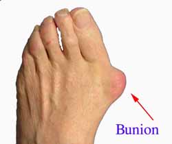
Surgical Treatment
If conservative treatment doesn't provide relief from your symptoms, you may need surgery. The goal of bunion surgery is to relieve discomfort by returning your toe to the correct position. There are a number of surgical procedures for bunions, and no one technique is best for every problem. Surgical procedures for bunions might involve removing the swollen tissue from around your big toe joint. Straightening your big toe by removing part of the bone. Realigning the long bone between the back part of your foot and your big toe, to straighten out the abnormal angle in your big toe joint. Joining the bones of your affected joint permanently. It's possible you may be able to walk on your foot immediately after a bunion procedure. However, full recovery can take weeks to months. To prevent a recurrence, you'll need to wear proper shoes after recovery. It's unlikely that you'll be able to wear narrower shoes after surgery. Surgery isn't recommended unless a bunion causes you frequent pain or interferes with your daily activities. Talk to your doctor about what you can expect after bunion surgery.
Overpronation Problems
Overview
When the ankles over-pronate, it throws everything out of alignment. The bones in the feet can actually shift over time, and the muscles and tendons in the leg and ankle will twist. This places repetitive stress on the ligaments, tendons, joints and muscles of the foot, which can cause injury. If your feet are over-pronating, you may notice your muscles feet unusually tired when you way or stand, or they may ache. But symptoms will vary by your weight, age and activity.
Causes
Congenital "Flat Feet" - an individual may be born with feet that lack an appropriately supportive arch thereby predisposing the individual to this foot condition. Excessive Weight (Obesity) Too much weight on the foot from either obesity or pregnancy may be a factor. Repetitive Impact walking on flat, hard surfaces continuously places unnatural stress on the foot arch.
Symptoms
Symptoms can manifest in many different ways. The associated conditions depend on the individual lifestyle of each patient. Here is a list of some of the conditions associated with over Pronation. Hallux Abducto Valgus (bunions). Hallux Rigidus (stiff 1st toe). Arch Pain. Heel Pain (plantar Facsitus). Metatarsalgia (ball of the foot pain). Ankle Sprains. Shin Splints. Achilles Tendonitis. Osteochondrosis. Knee Pain. Corns & Calluses. Flat Feet. Hammer Toes.
Diagnosis
Look at the wear on your shoes and especially running trainers; if you overpronate it's likely the inside of your shoe will be worn down (or seem crushed if they're soft shoes) from the extra strain.
Non Surgical Treatment
When you see the doctor, he or she will likely perform a complete examination of your feet and watch you walk. The doctor will need to take x-rays to determine the cause of your flat feet. In some cases, further imaging may be needed, especially if your symptoms are severe and sudden in nature. Once you are properly diagnosed, your doctor will create an appropriate treatment plan. There are several options to correct overpronation, such as orthotics. In many cases, overpronation can be treated with non-surgical methods and over-the-counter orthotics. In severe cases, however, custom-made orthotics may work better. Orthotics provide arch support and therefore prevent collapse of the arch with weight bearing. They are made of materials such as spongy rubber or hard plastic. Your doctor will also want to examine your footwear to ensure they fit properly and offer enough medial support. Extra support and stability can be achieved with footwear that has a firm heel counter. If you are experiencing pain, you should be able to use over-the-counter pain medications such as ibuprofen to relieve symptoms.
Prevention
Duck stance: Stand with your heels together and feet turned out. Tighten the buttock muscles, slightly tilt your pelvis forwards and try to rotate your legs outwards. You should feel your arches rising while you do this exercise.
Calf stretch:Stand facing a wall and place hands on it for support. Lean forwards until stretch is felt in the calves. Hold for 30 seconds. Bend at knees and hold for a further 30 seconds. Repeat 5 times.
Golf ball:While drawing your toes upwards towards your shins, roll a golf ball under the foot between 30 and 60 seconds. If you find a painful point, keep rolling the ball on that spot for 10 seconds.
Big toe push: Stand with your ankles in a neutral position (without rolling the foot inwards). Push down with your big toe but do not let the ankle roll inwards or the arch collapse. Hold for 5 seconds. Repeat 10 times. Build up to longer times and fewer repetitions.
Ankle strengthener: Place a ball between your foot and a wall. Sitting down and keeping your toes pointed upwards, press the outside of the foot against the ball, as though pushing it into the wall. Hold for 5 seconds and repeat 10 times.
Arch strengthener: Stand on one foot on the floor. The movements needed to remain balanced will strengthen the arch. When you are able to balance for 30 seconds, start doing this exercise using a wobble board.
Does Severs Disease Necessitate Surgical Treatment?
Overview
Sever's disease or calcaneal apophysitis, is the most common cause of heel pain in the growing athlete and is due to overuse and repetitive microtrauma of growth plates of the calcaneus in the heel. It occurs in children ages 7 to 15, with the majority of patients presenting between 10 and 12 years of age.
Causes
The condition is thought to result from repetitive microtrauma to growth plates of the calcaneus. Although some of the recent articles says, there is no evidence to support that weight and activity levels are risk factors for Sever's disease. High plantar foot pressures are associated with Sever's disease, although it is unclear whether they are a predisposing factor or a result of the condition. Gastrocnemius equinus may be a predisposing factor for Sever's disease.
Symptoms
Pain is usually felt at the back and side of the heel bone. Sometimes there may be pain at the bottom of the heel. The pain is usually relieved when the child is not active and becomes painful with sport. Squeezing the sides of the heel bone is often painful. Running and jumping make the symptoms worse. One or both heels can be affected. In more severe cases, the child may be limping.
Diagnosis
Sever condition is diagnosed by detecting the characteristic symptoms and signs above in the older children, particularly boys between 8 and 15 years of age. Sometimes X-ray testing can be helpful as it can occasionally demonstrate irregularity of the calcaneus bone at the point where the Achilles tendon attaches.
Non Surgical Treatment
If your child lets you know that his heels are hurting, schedule a doctor's appointment. Your family doctor may or may not refer you to a podiatrist. Treatment for Sever's Disease typically consists of one or more of the following steps. Reducing physical activity. Because Sever's Disease appears to be most common in athletic children, reducing exercise periods will relieve pressure on the heel bones, thereby reducing pain. Your doctor may recommend that your child take a complete break from athletic activity for a set amount of time. Icing the heel bones can help to lower both inflammation and pain levels. Use a cold pack or wrap ice in a towel and apply it to the heels. A new exercise regimen that involves simple stretches designed to lengthen the calf muscles and tendons. Your doctor may prescribe the use of orthotic shoe inserts that will assist your child in maintaining a good level of physical activity. HTP Heel Seats may be an excellent option and have been purchased by many parents as an effective aide for children suffering from Sever's Disease. Read about HTP Heel Seats here and ask your doctor if they are right for your child's unique case. In extreme cases, a doctor may recommend a plaster cast or boot, but typically only if other less cumbersome solutions fail to reduce pain. Some doctors may prescribe anti-inflammatory medications. Never give these to a child yourself, without first seeking a doctor's advice. Some medications carry the risk of serious side effects for children. Only give medications if specifically prescribed your child's physician.
Prevention
Can Calcaneal Apophysitis Be Prevented? The chances of a child developing heel pain can be reduced by avoiding obesity. Choosing well-constructed, supportive shoes that are appropriate for the child?s activity. Avoiding or limiting wearing of cleated athletic shoes. Avoiding activity beyond a child?s ability.
What Can Cause Posterior Tibial Tendon Dysfunction ?
Overview
Adult acquired flatfoot is one of the most common problems affecting the foot and ankle. Treatment ranges from nonsurgical methods, such as orthotics and braces to surgery. Your doctor will create a treatment plan for you based on what is causing your AAFD. 
Causes
Posterior tibial tendon dysfunction is the most common cause of acquired adult flatfoot. Sometimes this can be a result of specific trauma, but usually the tendon becomes injured from wear and tear over time. This is more prevalent in individuals with an inherited flat foot but excessive weight, age, and level of activity are also contributing factors.
Symptoms
Your feet tire easily or become painful with prolonged standing. It's difficult to move your heel or midfoot around, or to stand on your toes. Your foot aches, particularly in the heel or arch area, with swelling along the inner side. Pain in your feet reduces your ability to participate in sports. You've been diagnosed with rheumatoid arthritis; about half of all people with rheumatoid arthritis will develop a progressive flatfoot deformity.
Diagnosis
Diagnostic testing is often used to diagnose the condition and help determine the stage of the disease. The most common test done in the office setting are weightbearing X-rays of the foot and ankle. These assess joint alignment and osteoarthritis. If tendon tearing or rupture is suspected, the gold standard test would be MRI. The MRI is used to check the tendon, surrounding ligament structures and the midfoot and hindfoot joints. An MRI is essential if surgery is being considered.
Non surgical Treatment
What are the treatment options? In early stages an orthotic that caters for a medially deviated subtalar joint ac-cess. Examples of these are the RX skive, Medafeet MOSI device. Customised de-vices with a Kirby skive or MOSI adaptation will provide greater control than a prefabricated device. If the condition develops further a UCBL orthotic or an AFO (ankle foot orthotic) could be necessary for greater control. Various different forms of surgery are available depending upon the root cause of the issue and severity. 
Surgical Treatment
Until recently, operative treatment was indicated for most patients with stage 2 deformities. However, with the use of potentially effective nonoperative management , operative treatment is now indicated for those patients that have failed nonoperative management. The principles of operative treatment of stage 2 deformities include transferring another tendon to help serve the role of the dysfunctional posterior tibial tendon (usually the flexor hallucis longus is transferred). Restoring the shape and alignment of the foot. This moves the weight bearing axis back to the center of the ankle. Changing the shape of the foot can be achieved by one or more of the following procedures. Cutting the heel bone and shifting it to the inside (Medializing calcaneal osteotomy). Lateral column lengthening restores the arch and overall alignment of the foot. Medial column stabilization. This stiffens the ray of the big toe to better support the arch. Lengthening of the Achilles tendon or Gastrocnemius. This will allow the ankle to move adequately once the alignment of the foot is corrected. Stage 3 acquired adult flatfoot deformity is treated operatively with a hindfoot fusion (arthrodesis). This is done with either a double or triple arthrodesis - fusion of two or three of the joints in hindfoot through which the deformity occurs. It is important when a hindfoot arthrodesis is performed that it be done in such a way that the underlying foot deformity is corrected first. Simply fusing the hindfoot joints in place is no longer acceptable.